Week #34: May 10 - May 14

|
Make Up Week
This week our awesome anatomy will be a make up week. Take this opportunity to catch up on any missing assignments, or to double check any work. Triple check and read carefully to make sure you didn't miss any instructions. Awesome Anatomy Folders are due next Monday. Email Mr. Will if you have any questions. |
|
Week #33: May 3 - May 7
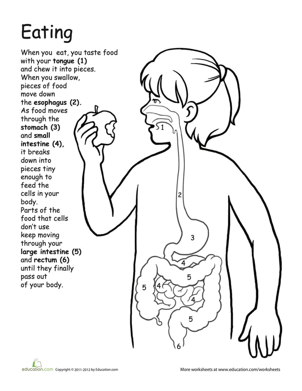
When you eat food, digestion starts in the mouth where your teeth and saliva begin the work of breaking down your food. When you swallow the broken down food pieces travel down the esophagus, the long tube that connects your mouth and your stomach. In th stomach the food is mixed with stomach acid and churned into a paste where it is sent to the small intestines. The small intestines are a misnomer, while the tube is narrow, the small intestines are nearly 25 feet long in a grown adult. The small intestines have lots of twists and turns, and these twists and turns are where most of your nutrients are absorbed. After the small intestines all you are left with only waste and liquids. The liquids are reabsorbed in the large intestines. The last stop in the process of digestion is the rectum where waste product is passed out of your system. Week #32: April 26 - April 30
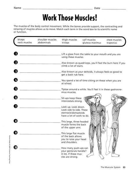
This page will ask you to think about how your muscles are used. Fill in the blank with the name of the muscles used to complete page 1. Page 2 is a matching game. Use a different color for each muscle and circle the action with that color. If you get stuck, you can perform the action and that will help you know the muscles you are using.Good Luck! Week #31: April 19 - April 23
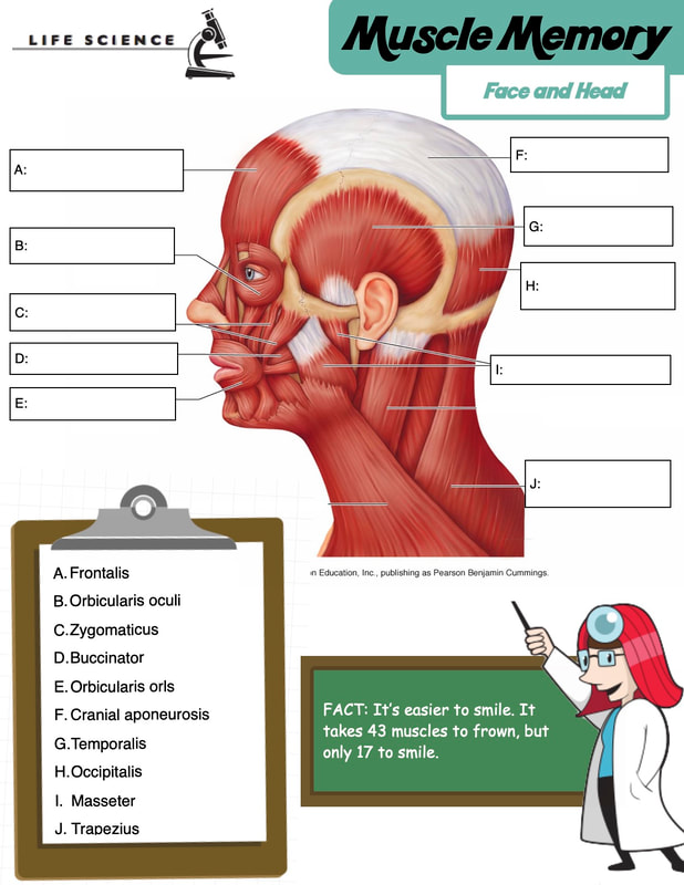
Most of these muscles exist around your facial bone structure. So if you look at the names of the muscles compared with the names of the bones you will see many similarities which will help you understand where the muscle is on the face. Remember it takes 43 muscles to frown and only 17 to smile. That's why it's always easier to smile and be happy. Week #30: April 12 - April 16
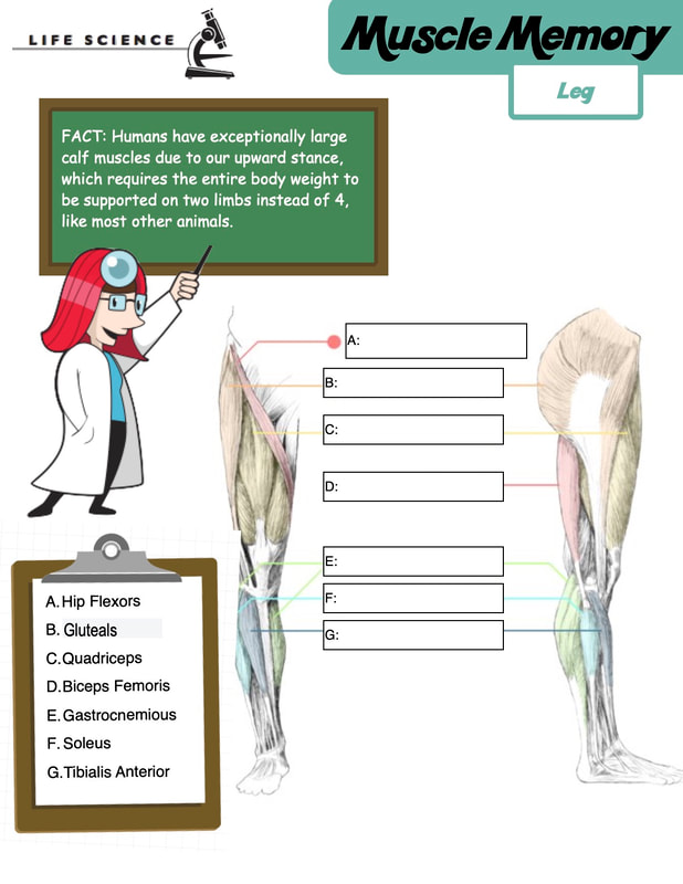
The quadriceps are the muscle group of the thigh. They help you straighten your leg. The biceps femoris, also known as your hamstrings, are the back side of your leg. they run from the back of your knee to just below your gluteals. Your gluteals are your sit muscles, and help you push when walking. The gastrocnemius and the soleus are your calf muscles. They are the muscles of your lower leg. |
|


Week #29: March 29 - April 2

|
Make Up Week
This week our awesome anatomy will be a make up week. Take this opportunity to catch up on any missing assignments, or to double check any work. Triple check and read carefully to make sure you didn't miss any instructions. Email Mr. Will if you have any questions. |
|
Week #28: March 22 - March 26
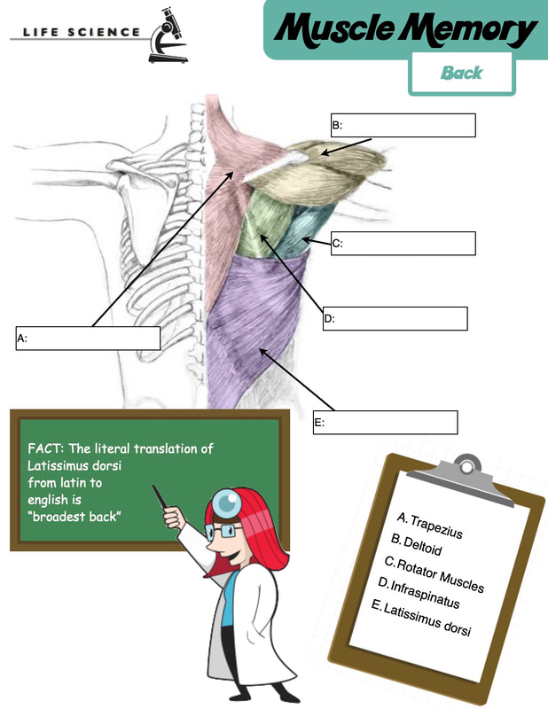
The largest muscle in your back is the Latissimus dorsi. Literally translated from latin to english Latissimus dorsi means broadest back. Your Trapezius is the shoulder muscle that connects to your neck and mid back. This muscle is often called the "I don't know muscle" because you use it to shrug your shoulders. The deltoids are your shoulder muscle that control your arm movements. At a certain point in their motion the rotator muscles will take over. The Infraspinatus is one of the under appreciated muscles of the back. It is also a shoulder rotator muscle, and it gets its name because it is below the other muscles and connects to your spine. Week #27: March 15 - March 9
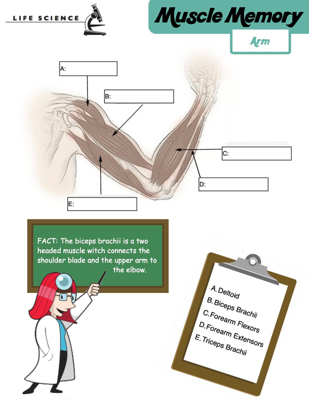
The biceps gets its name because it has two heads. Bi like bicycle meaning two. Likewise the ticeps have three heads. Tri like tricycle meaning three. The biceps and the triceps are called muscles of opposition because they work opposite each other. When bend your arm your bicep is contracting and your tricep is relaxing. When your straighten your arm your triceps are flexing, or contracting, and your bicep is relaxing. The same is true for the forearm. The forearm flexors bend your wrist inwards and your forearm extensor bends your wrist outwards. |
|


Week #26: March 8 - March 12

|
Make Up Week
This week our awesome anatomy will be a make up week. Take this opportunity to catch up on any missing assignments, or to double check any work. Triple check and read carefully to make sure you didn't miss any instructions. Email Mr. Will if you have any questions. |
|
Week #25: March 1 - March 5
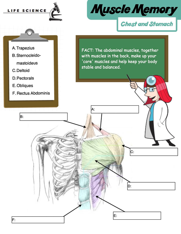
The Pectoral muscles are the muscles of your chest. These muscles are activated when you do pushups. The rectus abdominus muscles are the sit up muscles. they are the muscles many people refer to when they say you have a 6-pack. The Obliques are the side abdominal muscles. You uses the obliques when you twist from side to side. The deltoids are your shoulder muscles and help you lift your arms to the front and to the side. The Trapezius and Sternocleidomastoideus are your neck muscles. They help keep your head up. Week #24: February 22 - February 26
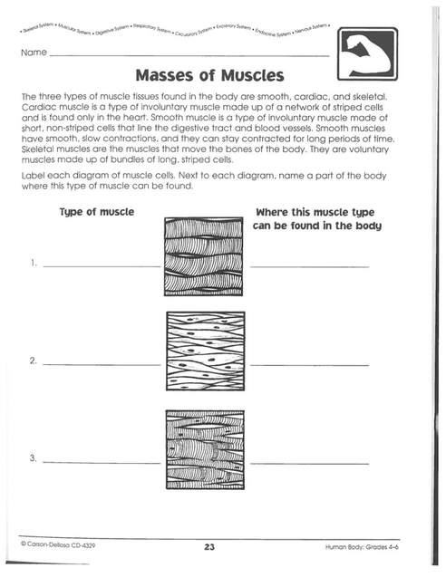
Cardiac muscle is a type of involuntary muscle made up of a network of stripped cells and is found only in the heart. Smooth muscle is a type of involuntary muscle made of short, non-striped class that line the digestive tract and blood vessels. Smooth muscles have smooth, slow contractions and can stay contracted for long periods of time. Skeletal muscles are the muscles that move the bones of the body. They are voluntary muscles made up of bundles of long, striped cells.. Week #23: February 8 - February 12
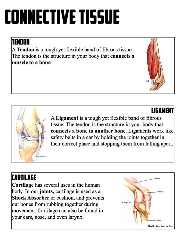
Tendons are the tough yet flexible band of fiberous tissue. Tendons are the structure of the body that connects muscles to bones. Ligaments are also a tough yet flexible band of fiberous tissue. Ligaments connect bone to bone. Ligaments work like safety belts in a car by holding the joints together in their correct place and stopping them from falling apart. Cartilage has several uses in the human body. In our joints cartilage is used as a shock absorber, cushioning and preventing our bones from rubbing together during movement. Cartilage can also be found in your ears, nose, and even your larynx. Week #22: February 1 - February 5
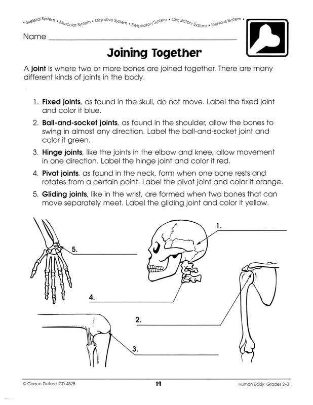
A fixed joint like the ones in the skull do not move Ball and socket joints can be found in the should and hip. These joints allow bones to swing in almost any direction. Like a door, hinge joints like the elbow and the knee allow movement in one direction. They open and close. Pivot Joints as found in the neck, form when one bone rests and rotates from a certain point. Gliding joints like the wrist are formed when two bones that can move separately meet. Week #21 : January 25 - January 29
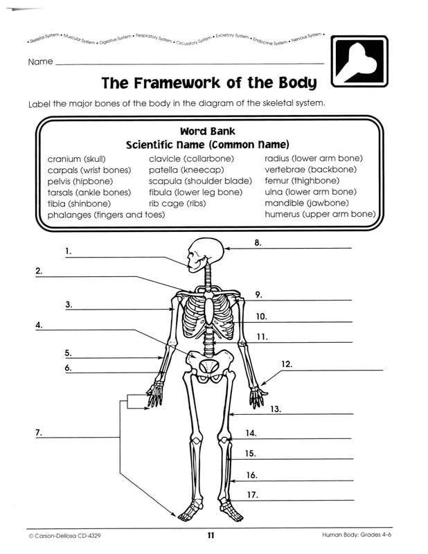
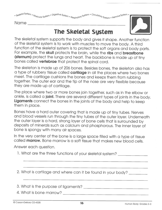
Bones are the part of the skeletal system that gives your body its size and shape. It also helps to protect vital organs in your body. Remember to follow all the directions on page 2 to get full credit, and that spelling doesn't count as much as neatness. Take your time and be sure to put both the scientific and common names for each bone on or near the line. Week #20 : January 19 - January 22
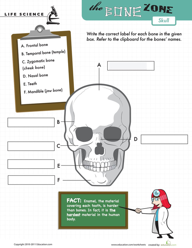
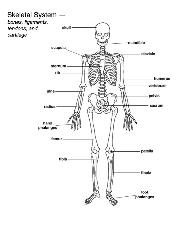
The bones of the skull include the Frontal Bone, Temporal Bone, Zygomatic Bones, Nasal Bone, The Mandible, and Teeth. The Frontal bone is the forehead bone. and the Temporal bone is the sides of your skull. Two other skull bones, the croronal bone and the occipital bone are not pictured but help make up the bulk of the skull. The Nasal Bone and the Zygomatic bones are your facial bones. They are what give your face its shape. The Mandible is the jaw bone and makes up the only moveable joint in the skull. Lastly we have Teeth. Teeth are the only bones in your body exposed to the world, because of that they are coated in a hard material called enamel that keeps them from breaking. |
|


Week #19: January 11 - January 15

|
2021 Goal Setting Due
Be sure to complete at least 1 smart goal from the worksheet below and share it via Seesaw or Canvas with me. If you share your goal with me I can help you accomplish your goal and celebrate when you achieve success. |
|
Week #18: January 4 - January 8
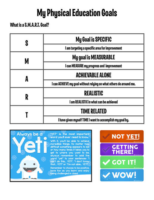
A SMART goal is: Specific, Measurable, Achievable, Realistic & Relevant, and lastly Time Bound. Use the sheet to the right to set three of your own goals. Start at Not Yet and work your way to Wow! Remember to always be a YETI! It is important to have a growth mindset when it comes to P.E. When you struggle to do something instead of saying "I can't," try "I can't YET." |

|

Week #17 December 14 - December 18

|
REMEMBER TO TURN IN YOUR PORTFOLIOS FOR CREDIT!
This week we are turning in our completed portfolios. During the break, I will be grading and assessing the portfolios for completion. These portfolios can help your grade tremendously. When you get your portfolios back they will be filled with more new sheets as well, so remember to turn them in so that you can keep learning all the awesome things about our anatomy. |
|
Week #16: December 7 - December 11
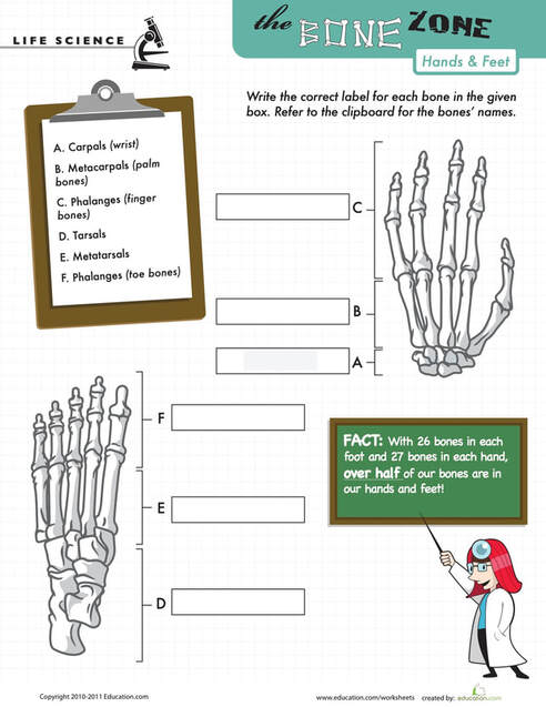
The wrist bones are scientifically known as Carpals. These small round bones each have an individual name. They are small in order to allow maximum movement. The Heel bones are known as Tarsals. Unlike the Carpals the Tarsals are long and flat. This prevents range of motion but increases balance and support. Metacarpals are the bones of the hands. Similarly the bones of the foot are called Metatarsals. These bones are flexible and allow you to curl your hands and feet. Both your Fingers and your toes are scientifically known by the same name: Phalanges. |
|

Week #15: November 30 - December 4

|
Final Make Up Week
This week we will be virtual, and this will be your final week to complete any missing assignments before we turn them in before Winter Break. I will be reviewing and grading your portfolios during break and adding new content for the weeks between now and the end of the year. Be sure to double or even triple check your work to make sure you didn't miss anything. |
Week #14: November 23 - November 24

|
Thanksgiving Week
This week I will be on vacation. Please use this time to catch up on any missing Awesome Anatomy Assignments. Awesome Anatomy Portfolios will be collected and graded the week after Thanksgiving. Have a safe and enjoyable holiday. HAPPY THANKSGIVING! |
Week #13: November 16 - November 20

|
Make Up Week
This week our awesome anatomy will be a make up week. Take this opportunity to catch up on any missing assignments, or to double check any work. Triple check and read carefully to make sure you didn't miss any instructions. Email Mr. Will if you have any questions. |
|
Week #12: November 9 - November 13
The upper leg bone is called the femur. The femur is the largest and longest single bone in your body. It makes up 1/4 of your total height. The patella is commonly called the knee cap and helps protect the joint between the upper leg and the shin bones. The two shin bones are called the tibia and fibula. The tarsals are your ankle and heel bones The metatarsals are the bones of the feet, and yYour toes just like your fingers are phalanges. The structure of your arm is very similar to the structure of your leg. Can you name 3 similarities and 3 differences? Week #11: November 2 - November 6
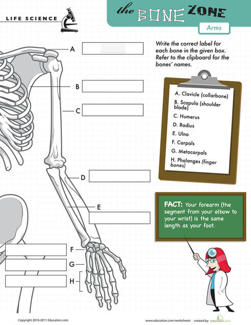
The Upper arm bone is called the humerus and connects with the shoulder. It stretches halfway down your arm.
There are two lower arm bones. The thumb side bone is called the radius, and the pinky side lower arm bone is called the ulna. Next come the wrist bones scientifically called carpals. The metacarpals are your interior hand bones. Lastly comes our phalanges, also known as the finger bones. Week #10: October 26 - October 30
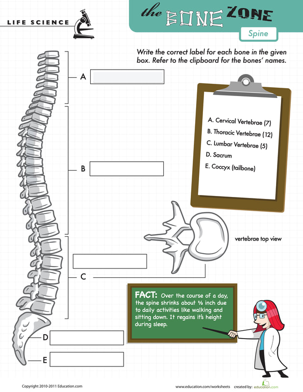
Cervical Vertebra are the first 7 vertebrae as you go down from your head. These are small vertebrae allowing for the most movement.
Thoracic Vertebrae are the vertebrae that are attached to the rib cage and make up the next 12 vertebrae. Lumbar Vertebrae make up the lower part of your back bone. these vertebrae are the largest vertebrae in the body because they have to support more weight that the others. You Sacrum and is attached to the pelvis and is part of your pelvic girdle, and The Coccyx is your tailbone. Week #9: October 19 - October 23
The spine is your backbone and stretches the length of your torso.
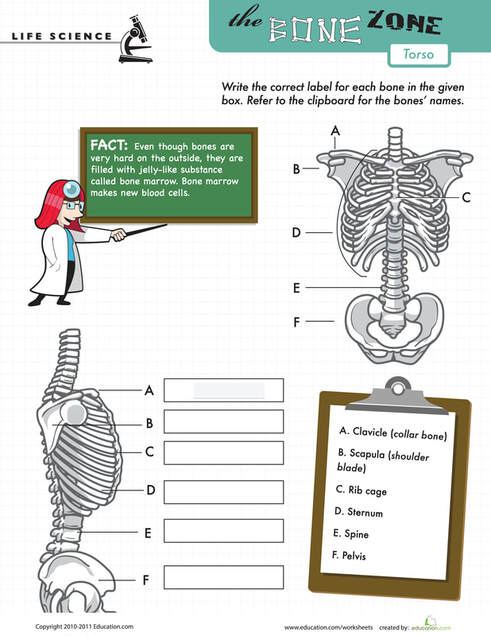
Your Scapula is a flat bone that is commonly called your shoulder blade. Your Clavicle is the cylindrical bone commonly known as your collar bone. Your Ribs and Sternum are fused together and create the Rib Cage. Your Pelvis is your hip bone. You can remember this because "Elvis Shook his Pelvis" Week #8: October 13 - October 16
Bones have a hard outer covering that is made up of tiny tubes. Nerves and blood vessels run through the tiny tubes of the outer layers.
Underneath the outer layer is a hard, strong layer of bone cells that is surrounded by deposits of minerals such as calcium and phosphorus. The inner layer of bone is spongy with many air spaces. In the very center of the spongy bone layer is a tissue called Bone Marrow. Bone Marrow is where blood cells are made. Week #7: October 5 - October 9
Second the parts of the skeletal system is used to protect vital organs such as your spinal cord, lungs, heart, kidneys and brain.
The primary components of the skeletal system are Bones, Ligaments, Tendons and Cartilidge. Bones are the hard structures that make up your skeleton. Ligaments connect bones to other bones. Tendons connect muscles to bones, and Cartilidge is a cushioning material that exists between bones and can also be found in your ears and nose. |

|


Week #6: September 28 - October 2

|
Make Up Week
This week our awesome anatomy will be a make up week. Take this opportunity to catch up on any missing assignments, or to double check any work. Triple check and read carefully to make sure you didn't miss any instructions. Email Mr. Will if you have any questions. |
|
Week #5: September 21 - September 25
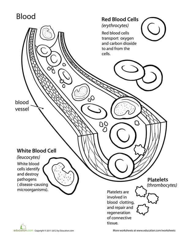
Arteries carry the blood away from your heart, veins carry the blood back towards your heart. Capillaries are the smallest blood vessels in the body and connect arteries to veins.
Your Blood consists of Blood Cells and Blood Plasma. Blood Plasma is the liquid substance that blood is made of. Red blood cells transport oxygen and carbon dioxide to and from the body. White blood cells are your bodies disease fighting cells. They are part of your immune system. Platelets help your blood clot and repair damaged tissue. Week #4: September 14 - September 18
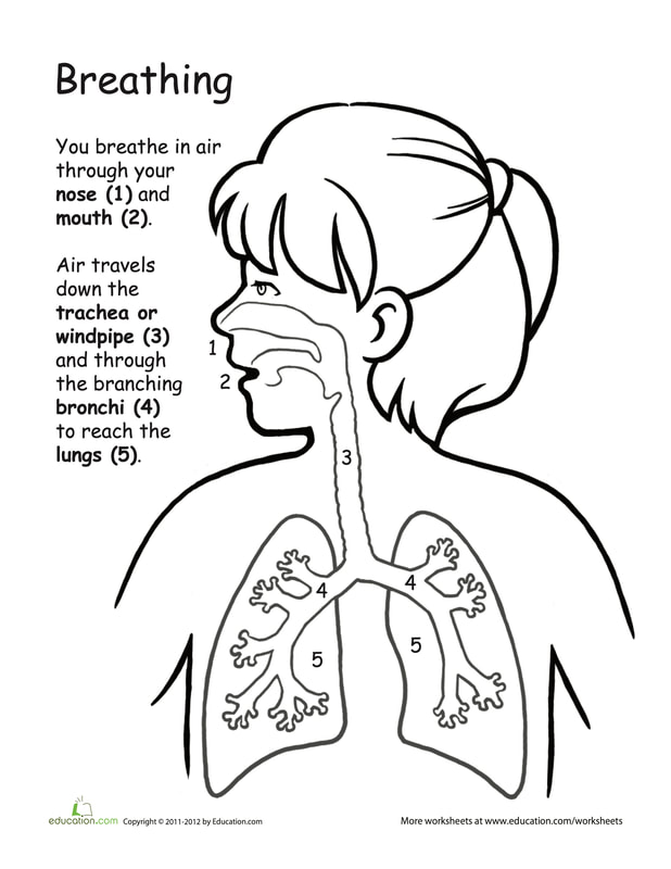
You breathe in air through your nose and mouth.
Air travels down the trachea or windpipe. Do not confuse your windpipe and your esophagus they are two separate tubes. The trachea will then divide into two separate smaller tubes known as bronchi tubes. at the end of these are small air sacks called alveoli. The lungs are less like balloons and more like spongy tissue created by the alveoli. Week #3: September 8 - September 11
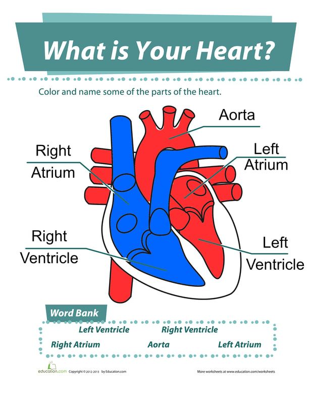
The right side of the heart fills with non oxygenated blood from the body. It then pumps to the lungs.
The left side of the heart receives oxygen rich blood from the lungs. The left ventricle pumps blood out through the Aorta, the largest artery in the body. The Aorta may be an Artery but it is also considered part of the heart. Week #2: August 31 - September 4
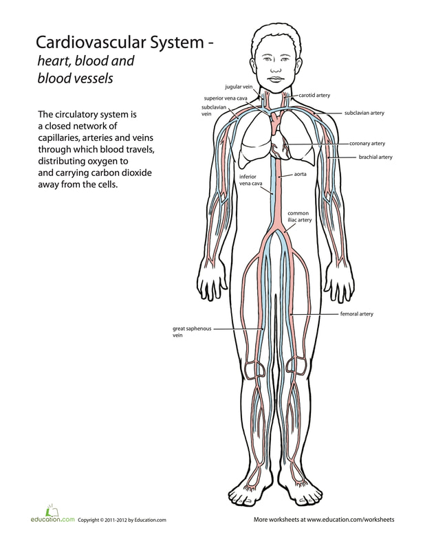
The Heart is the pump that pushes blood throughout the body.
The Lungs are your body's way of obtaining oxygen. Your lungs also help your body remove carbon dioxide. The Blood Vessels are your arteries and veins. Arteries and veins are the way that your body uses to transport nutrients to your body's cells. Arteries cary blood away from your heart, and veins cary blood back towards your heart. Week #1: August 25 - 28
A SMART goal is: Specific, Measurable, Achievable, Realistic & Relevant, and lastly Time Bound.
Use the sheet to the right to set three of your own goals. Start at Not Yet and work your way to Wow! Remember to always be a YETI! It is important to have a growth mindset when it comes to P.E. When you struggle to do something instead of saying "I can't," try "I can't YET." |
|